Home
/ Animal Cell Seen Through Electron Microscope / Fixed Cells Useful Notes On The Structure Of Fixed Cells - It can be used to view dead and living samples and can maximize these samples up to one thousand times their actual size.
Animal Cell Seen Through Electron Microscope / Fixed Cells Useful Notes On The Structure Of Fixed Cells - It can be used to view dead and living samples and can maximize these samples up to one thousand times their actual size.
Animal Cell Seen Through Electron Microscope / Fixed Cells Useful Notes On The Structure Of Fixed Cells - It can be used to view dead and living samples and can maximize these samples up to one thousand times their actual size.. Sep 29, 2020 · animal cell as shown above. Here's a diagram of a plant cell: Image:animal cell seen under electron microscope. The electron microscope is more powerful than the light microscope. Below the basic structure is shown in the same animal cell, on the left viewed with the light microscope, and on the right with the transmission electron.
Resolving power is the ability to distinguish between separate things which are close to each other. Here's a diagram of a plant cell: Introduction to the lesson now that we have the basic understanding that all living things are composed of cells, we begin to move further and in this lesson we will learn how people study cells. Now the first thing to point out when looking at images under an electron microscope is the scale. It uses a beam of electrons to illuminate the specimen instead of light as in the case of light microscope.
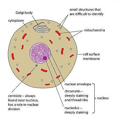
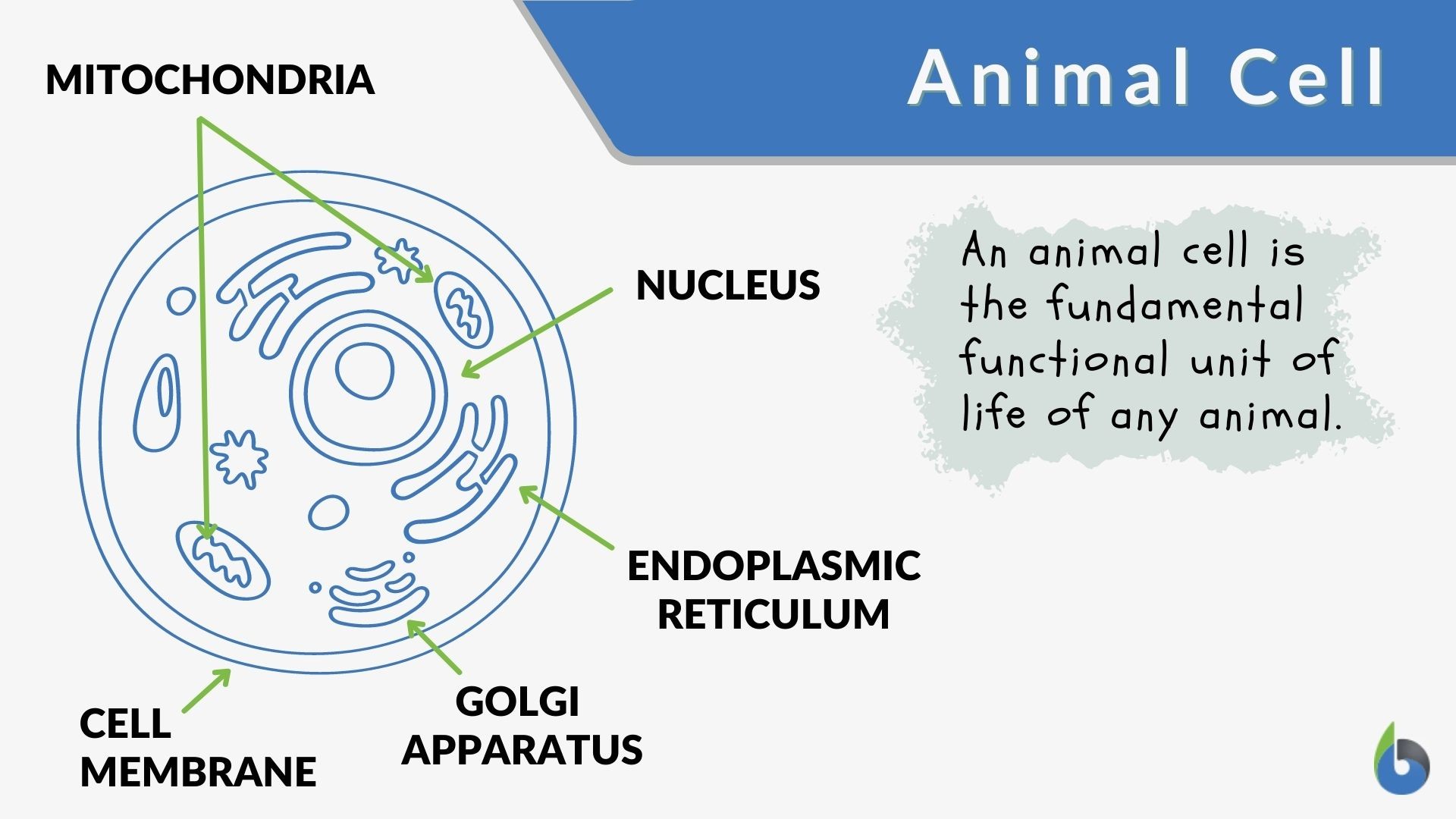
Topic Labeling Animal And Plant Cells Under The from slidetodoc.com Resolving power is the ability to distinguish between separate things which are close to each other. 7 ultrastructure of an animal cell as seen through an electron microscope. Animal cells have a basic structure. Introduction to the lesson now that we have the basic understanding that all living things are composed of cells, we begin to move further and in this lesson we will learn how people study cells. Structure and function of bacterial cells. Here's a diagram of a plant cell: More images for animal cell seen through electron microscope » Under the microscope, animal cells appear different based on the type of the cell.
More images for animal cell seen through electron microscope »
Image:animal cell seen under electron microscope. Resolving power is the ability to distinguish between separate things which are close to each other. Draw and label an animal cell as seen under an electron microscope. For example, something that you draw as 3cm long, may in fact be 10, 000 times smaller in real life. Jul 09, 2014 · a typical animal cell (as seen in an electron microscope) medical images for powerpoint 1. Typical animal cell pinocytotic vesicle lysosome golgi vesicles golgi vesicles rough er (endoplasmic reticulum) smooth er (no ribosomes) cell (plasma) membrane mitochondrion golgi apparatus nucleolus nucleus centrioles (2) each composed of 9 microtubule triplets microtubules cytoplasm ribosome Animal and plant cell under electron microscope. Difference between animal and plant cell. It can be used to view dead and living samples and can maximize these samples up to one thousand times their actual size. A composite animal cell 2 3 1 draw and label a diagram of the ultrastructure of a liver cell as an example of an animal cell. We trust you enjoy it! Here's a diagram of a plant cell: Asked nov 28, 2017 in class.
Aug 01, 2021 · here's a diagram of a plant cell: Plant cell as shown above. Introduction to the lesson now that we have the basic understanding that all living things are composed of cells, we begin to move further and in this lesson we will learn how people study cells. It can be used to view dead and living samples and can maximize these samples up to one thousand times their actual size. For example, something that you draw as 3cm long, may in fact be 10, 000 times smaller in real life.
Animal Cell Structure And Organelles With Their Functions Jotscroll from www.jotscroll.com Typical animal cell pinocytotic vesicle lysosome golgi vesicles golgi vesicles rough er (endoplasmic reticulum) smooth er (no ribosomes) cell (plasma) membrane mitochondrion golgi apparatus nucleolus nucleus centrioles (2) each composed of 9 microtubule triplets microtubules cytoplasm ribosome Animal and plant cell under electron microscope. Image:animal cell seen under electron microscope. Difference between animal and plant cell. Here's a diagram of a plant cell: Label the parts that carry on the function of respiration, secretion, protein synthesis, transport of material 2 May 29, 2016 · q14 draw a large diagram of an animal cell as seen through an electron microscope. Electron microscope can magnify an object up to 500,000 times.
Image:animal cell seen under electron microscope.
Draw and label an animal cell as seen under an electron microscope. Below the basic structure is shown in the same animal cell, on the left viewed with the light microscope, and on the right with the transmission electron. Introduction to the lesson now that we have the basic understanding that all living things are composed of cells, we begin to move further and in this lesson we will learn how people study cells. Difference between animal and plant cell. Here's a photo of a plant cell under an electron microscope. Aug 01, 2021 · here's a diagram of a plant cell: Jul 09, 2014 · a typical animal cell (as seen in an electron microscope) medical images for powerpoint 1. Here's a diagram of a plant cell: Image:animal cell seen under electron microscope. Aug 11, 2021 · image:plant cell seen under electron microscope. Now the first thing to point out when looking at images under an electron microscope is the scale. It uses a beam of electrons to illuminate the specimen instead of light as in the case of light microscope. The electron microscope is more powerful than the light microscope.
We trust you enjoy it! Image:animal cell seen under electron microscope. Introduction to the lesson now that we have the basic understanding that all living things are composed of cells, we begin to move further and in this lesson we will learn how people study cells. Animal cells have a basic structure. Asked nov 28, 2017 in class.
Animal Cell Definition And Examples Biology Online Dictionary from www.biologyonline.com Here's a diagram of a plant cell: Here's a photo of a plant cell under an electron microscope. Label the parts that carry on the function of respiration, secretion, protein synthesis, transport of material 2 Resolving power is the ability to distinguish between separate things which are close to each other. 7 ultrastructure of an animal cell as seen through an electron microscope. Below the basic structure is shown in the same animal cell, on the left viewed with the light microscope, and on the right with the transmission electron. Secretly, they're all microscope freaks. Here's a diagram of a plant cell:
Jul 09, 2014 · a typical animal cell (as seen in an electron microscope) medical images for powerpoint 1.
The cell as seen under the electron microscope. Under the microscope, animal cells appear different based on the type of the cell. More images for animal cell seen through electron microscope » The electron microscope is more powerful than the light microscope. Electron microscope can magnify an object up to 500,000 times. Difference between animal and plant cell. For example, something that you draw as 3cm long, may in fact be 10, 000 times smaller in real life. It uses a beam of electrons to illuminate the specimen instead of light as in the case of light microscope. Image:animal cell seen under electron microscope. We trust you enjoy it! May 29, 2016 · q14 draw a large diagram of an animal cell as seen through an electron microscope. Typical animal cell pinocytotic vesicle lysosome golgi vesicles golgi vesicles rough er (endoplasmic reticulum) smooth er (no ribosomes) cell (plasma) membrane mitochondrion golgi apparatus nucleolus nucleus centrioles (2) each composed of 9 microtubule triplets microtubules cytoplasm ribosome 7 ultrastructure of an animal cell as seen through an electron microscope.
Post a Comment
for "Animal Cell Seen Through Electron Microscope / Fixed Cells Useful Notes On The Structure Of Fixed Cells - It can be used to view dead and living samples and can maximize these samples up to one thousand times their actual size."


Post a Comment for "Animal Cell Seen Through Electron Microscope / Fixed Cells Useful Notes On The Structure Of Fixed Cells - It can be used to view dead and living samples and can maximize these samples up to one thousand times their actual size."